Medical Terminology for Cancer
© Copyright 1996-2013
6: The Skeletal System (Bones)
Contents
 Functions of the skeletal system The skeleton Parts of a bone Types of bone Bones and growth Roots, suffixes, and prefixes Cancer Focus Related Abbreviations and Acronyms Further Resources
Functions of the skeletal system
There are about 206 bones in the human body, they have the function of protecting and preserving the shape of soft tissues. The skeleton provides a framework for the muscles, it controls and directs internal pressure and provides stability anchoring points for other soft tissues. There are a wide variety of bones/bony tissues adapted for specific functions to aid locomotion and support, bones are moved by the skeletal muscles. In addition the skeletal system stores and produces blood cells in the bone marrow.The skeleton
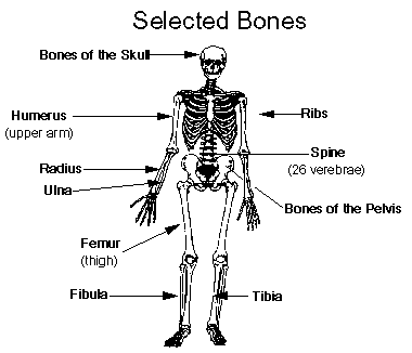
It is not the aim of this guide to catalogue each bone, but the following may be useful:
- Thorax
- the bones of the thorax (ribs, sternum and thoracic vertebrae) form a cage which protects many of the body's vital organs.
- The Axial skeleton
- This is the main body including the pelvis, thorax, and skull (excluding the arms and legs).
- The Proximal skeleton
- The femur and humerus (ie the bones proximal to the Axial skeleton)
- The Distal skeleton
- The lower legs (tibia, fibula, and feet bones) and lower arm (radius, ulna, and bones of the hand). The Proximal and Distal skeleton are sometimes collectively referred to as bones of the extremities.
- The spine
- The spine is divided into 5 main areas and each bone (verebrae) has a letter and number:
- Cervical vetebrae C1 - C7
- the neck region. C1 is the upper most vertebrae.
- Thoracic vertabrae T1 - T12
- vertebrae of the upper body (thorax)
- Lumbar vertebrae L1 - L5
- vertebrae of the lower back
- Bones of the sacrum S1 - S5
- vertebra within the pelvic girdle. These bones fuse together between ages 16 and 18.
- The coccyx Co1 - Co4
- The lower tip of the spine. These bones fuse together between ages 20 to 30.
Parts of a bone
- Diaphysis
- The long shaft of the bone.
- Epiphysis
- The knob like end of the bone, often contains red marrow (blood cells).
- Metaphysis
- Region where the diaphysis joins the epiphysis, important in bone growth.
- Medullary
- Marrow cavity inside the bone. Contains yellow marrow (fat cells).
- Foramina
- Tiny canals in the bone through which blood and lymph vessels connect to the medullary.
- Cartilage
- Tough connective tissue covering the ends of the bone. The cartilage reduce friction and acts as a shock absorber.
- Ligament
- Fibrous tissue that connects bones or cartilage to strengthen and support joints.
The end of the bones are often refered to by the Proximal end (towards the main body) or the Distal end (away from the main body), e.g. the proximal femur is the top end of the thigh bone.
Types of bone
- Compact bone
- Compact bone is dense and hard, especially the outer layer of the bone
- Spongy bone
- Made up of a lattice work of bone, the spaces are filled with red marrow which produce blood cells.
Classification of bones by shape:
- Long bones
- Slightly curved for STRENGTH with long narrow shafts with knobbly ends (especially found in arms and legs e.g. femur).
- Short bones
- Tend to be spongy e.g. wrists, fingers, toes and ankles.
- Flat bones
- Plate like and highly PROTECTIVE e.g. bones of the skull protect the brain.
- Irregular bones
- e.g. vertebrae (spine)
Bones and growth
Ossification is the gradual conversion of cartilage or other tissue into bone. At birth ossification is not complete, there are still may membrane filled spaces in the skull, these are called fontanels or "soft spots". Most bone growth occurs during childhood, and ossification of most bones is usually complete by age 25. The 5 bones of the sacrum fuse together from ages 18 to 25. When all bone growth is complete the body is said to be skeletally mature.Roots, suffixes, and prefixes
Most medical terms are comprised of a root word plus a suffix (word ending) and/or a prefix (beginning of the word). Here are some examples related to the Skeletal System. For more details see Chapter 4: Understanding the Components of Medical Terminology
| component | meaning | example |
| ARTHR- | joint | arthritis = inflammation of the bone |
| CHONDR- | cartilage | chondrocyte = a cartilage cell |
| COST- | rib | costalgia = pain in the ribs |
| OSTEO- | bone | osteosarcoma = a type of bone tumour |
| SCOLIO- | curved / crooked | scoliosis = curvature of the spine |
| -LYSIS | disintegration | osteomyelitis = inflammation of the bone |
| -OSIS | disease | osteoporosis = reduced bone mass-fracture prone |
| -TOMY | incision into | thoracotomy = incision into chest/thorax |
Cancer Focus
- Osteogenic Sarcoma
- Osteogenic Sarcoma (osteosarcoma) is a bone forming cancer. It is the most frequent type of bone tumour and is most common between the ages of 15 to 25. Over 90% of tumours are located in the metaphysis (the growing ends of the bone), the most common sites
are the long bones of the legs. Most tumours are solitary, around 2% are multifocal (2 or more bones). It is known that osteosarcoma can be radiation induced. Osteosarcomas vary greatly in radiological and pathological features and therefore needs careful
diagnosis to differentiate this from other bone tumours. Most are high grade intramedullary osteosarcomas, about 5% are low grade lesions, some are secondary osteosarcomas (for example those caused by radiation therapy).
 Internet Resources for Osteosarcoma
- Ewing's Sarcoma
- Ewing's sarcoma is most common in children and young adults. The most frequent sites are the pelvis, femur, tibia, and fibula, around a fifth of patients have metastases at diagnosis usually in the lungs or other other bones. Ewing's tumours are more
frequently found in the diaphysis (mid-shaft) part of the bone. Ewing's sarcoma can sometimes be restricted to soft tissue (Extraosseos Ewing's sarcoma). There is a spectrum of pathology ranging from 'classical' Ewing's which are negative for neural
markers; to PNET (peripheral neuroectodermal tumours) which are strongly positive.
Internet Resources for Ewing's Sarcoma
- Chrondrosarcoma
- Chondrosarcoma is a cancer arising in cartilage cells, it occurs mostly in adults, it is rare in those aged under 20 with 70% of cases occurring between ages 50-75. Rare sub-types include
mesenchymal chondrosarcoma which is more common in those aged under 40; Clear cell chondrosarcoma (around 2% of cases); and Dedifferenting chondrosarcoma (a rare tumour which transforms from low grade to a high grade sarcoma).
Internet Resources for Chondrosarcoma
- Other Primary Bone Tumours
- Malignant Fibrous Histiocytoma (MFH) account for 10% of bone tumours, they arise from histiocytes that have fibroblastic potential.
Chondoma is rare occurring mostly between ages 30 -70. This is a low grade malignancy which is arises from the remnants of the notochord (cells in the embryo which cause the formation of cartilage).
Fibrosarcoma and desmoid tumours vary in malignant potential.
Giant cell tumours some are benign, others may be malignant e.g. giant cell osteosarcoma, giant cell fibrosarcoma.
Neurogenious tumours of bone These include neuroepithelioma and malignant schwannoma.
Intraosseous liposarcoma is very rare. Liposarcoma arises from fat cells, these tumours are common in muscle but in rare cases are found in bone. Similarly extraosseous osteosarcoma (osteosarcoma found in soft tissue) is very rare with less than 300 cases reported world wide. - Bone sarcomas associated with Paget's disease
- Paget's disease is the most common bone disorder characterised by irregular thickening and softening of the bones. The disease is more common after the age of 40, and is frequent in those of European descent but rare in Asians. These is an association with this (non malignant) disease and bone cancer, up to 10% of those with Paget's disease will have a 'sarcomatous transformation' of affected bones giving rise to bone sarcoma. This may be osteosarcoma, fibrosarcoma, chondrosarcoma, or other bone sarcomas.
- Pathologic Fracture
- caused by a disease, growth of the tumour may cause stress leading to a fracture.
- Scintigraphy
- Scintigraphy (bone scan) - e.g. used to see if a cancer has metastasised to the bone. Primary bone cancers are relatively rare, but many other types of cancer can metastasise to the bone.
Related Abbreviations and Acronyms
| C1 - C7 | Cervical vertebrae (spine eg. C7 = seventh cervical vertebra) |
| EOI | European Osteosarcoma Intergroup |
| GCT | Giant Cell Tumour Context: bone tumours |
| IESS | Intergroup Ewing's Sarcoma Study (USA) |
| L1 - L5 | Lumbar vertebrae 1 - 5 (spine eg. L1 = 1st lumbar vertebra) |
| OS | Osteogenic sarcoma (context bone tumours) |
| PNET | Peripheral neuroectodermal tumour Context: Bone tumours - see Ewing's tumour |
| T1 - T12 | Thoracic vertebrae 1-12 (spine eg. T10 = tenth thoracic vertibra) |
Further Resources (3 links)
WebAnatomy, University of Minnesota
Test your anatomy knowledge with these interactive questions. Includes different question types and answers.
SEER, National Cancer Institute
Part of a SEER training module for cancer registry staff.
 The Skeletal System
Paul Andersen
Paul Andersen describes the important features of the skeletal system. He starts by comparing and contrasting endoskeletons and exoskeletons. He then explains how the human skeleton provides support, movement, storage, blood production and homeostasis.


This guide by Simon Cotterill
First created 4th March 1996
Last modified: 1st February 2014
 CancerIndex