MYCN

Gene Summary

Gene:MYCN; MYCN proto-oncogene, bHLH transcription factor
Aliases: NMYC, ODED, MODED, N-myc, bHLHe37
Location:2p24.3
Summary:This gene is a member of the MYC family and encodes a protein with a basic helix-loop-helix (bHLH) domain. This protein is located in the nucleus and must dimerize with another bHLH protein in order to bind DNA. Amplification of this gene is associated with a variety of tumors, most notably neuroblastomas. Multiple alternatively spliced transcript variants encoding different isoforms have been found for this gene. [provided by RefSeq, Jun 2014]
Databases:OMIM, HGNC, Ensembl, GeneCard, Gene
Protein:N-myc proto-oncogene protein
Source:NCBIAccessed: 01 September, 2019

Ontology:

What does this gene/protein do?
Show (17)

Cancer Overview

MYCN is over-expressed in a number of different types of cancer, most notably neuroblastoma, but also inclusing rhabdomyosarcoma, medulloblastoma, astrocytoma, Wilms' tumour, and small cell lung cancer. In neuroblastoma MYCN amplification is an established indicator of poor-prognosis. MYCN belongs to a family of similar transcription factors (including C-MYC).

FISH analysis on neuroblastoma from Pistoia et al., Front Oncol. 2012; 2: 174. License: CC BY 3.0
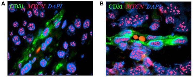
Neuroblastoma-derived endothelial micro-vessels. (A) Immunofluorescence and fluorescent in situ hybridization analysis of NB tumor section highlights CD31+ endothelial micro-vessel (green) carrying MYCN amplification (multiple red signals). (B) Two RBCs are in the open lumen of the NB-derived endothelial micro-vessel. Nuclei are stained with DAPI (blue), ×100.
Image from: from Pistoia et al, MYCN: from oncoprotein to tumor-associated antigen Front Oncol. 2012; 2: 174. License: CC BY 3.0.


Research Indicators

Publications Per Year (1994-2019)
Graph generated 01 September 2019 using data from PubMed using criteria.

Literature Analysis

Mouse over the terms for more detail; many indicate links which you can click for dedicated pages about the topic.

Tag cloud generated 01 September, 2019 using data from PubMed, MeSH and CancerIndex

Specific Cancers (9)

Data table showing topics related to specific cancers and associated disorders. Scope includes mutations and abnormal protein expression.

Entity Topic PubMed Papers
NeuroblastomaMYCN amplification in NeuroblastomaPrognostic
Amplification (duplicate copies) of the MYCN gene (also referred to as N-MYC) is established as an adverse prognostic factor in neuroblastoma. The amplicon (material co-amplified with MYCN) varies between patients, but can include the DDX1 gene. MYCN amplification is also correlates with 1p36 deletion and gain of chromosome 17q. MYCN amplified cell lines also overexpress ID2.
View Publications1364
RetinoblastomaMYCN Amplification in Retinoblastoma
There have been a number of reports of MYCN amplification in retinoblastoma cell lines and primary tumours. Doz et al. (1996) found only 1 of 45 primary retinoblastomas studies exhibited MYCN amplification while Kim et al. (1999) found 6 of 33 primary tumours were amplified. The latter study also suggested that MYCN amplified tumours have higher proliferation levels than non-amplified tumours.
View Publications113
RhabdomyosarcomaMYCN Amplification in Rhabdomyosarcoma
MYCN gene amplification occurs in a subset patients with rhabdomyosarcoma. It has been reported in 40-60% of patients with alveolar rhabdomyosarcoma, but infrequently occurs in patients with embyronal rhabdomyosarcoma. Some studies suggest that MYCN amplification in may be associated with a worse prognosis in rhabdomyosarcoma, however, findings have generally been based on small numbers.
View Publications61
Lung CancerMYCN in Lung Cancer View Publications51
MedulloblastomaMYCN Amplification in Medulloblastoma View Publications28
OsteosarcomaMYCN Amplification in Osteosarcoma View Publications13
NeuroblastomaABCC1 (MRP1) Overexpression in Neuroblastoma
Overexpression of MRP1 has been reported to have prognostic significance in neuroblastoma (Haber, 2006). There is also evidence that MRP1 is a target of MYCN (Blanc, 2003 & Manohar, 2004), frequently amplified in neuroblastoma, and involved in the development of multidrug resistance.
View Publications10
Wilms TumourMYCN amplification in Wilms Tumor
Williams et al (2010) reported MYCN amplification in 9% of cases in a SIOP study of over 100 Wilms tumor patients. This study also found that FBXW7 was mutated or deleted in approximately 4% of cases and the authors note that MYCN is a target of FBXW7-mediated ubiquitination and degradation - suggesting a common pathway is dysregulated by different mechanisms in various Wilms tumor subtypes.
View Publications10
Breast CancerMYCN and Breast Cancer View Publications4

Note: list is not exhaustive. Number of papers are based on searches of PubMed (click on topic title for arbitrary criteria used).

Latest Publications: MYCN (cancer-related)

Bishayee K, Habib K, Sadra A, Huh SO
Targeting the Difficult-to-Drug CD71 and MYCN with Gambogic Acid and Vorinostat in a Class of Neuroblastomas.
Cell Physiol Biochem. 2019; 53(1):258-280 [PubMed] Related Publications
BACKGROUND/AIMS: Although neuroblastoma is a heterogeneous cancer, a substantial portion overexpresses CD71 (transferrin receptor 1) and MYCN. This study provides a mechanistically driven rationale for a combination therapy targeting neuroblastomas that doubly overexpress or have amplified CD71 and MYCN. For this subset, CD71 was targeted by its natural ligand, gambogic acid (GA), and MYCN was targeted with an HDAC inhibitor, vorinostat. A combination of GA and vorinostat was then tested for efficacy in cancer and non-cancer cells.
METHODS: Microarray analysis of cohorts of neuroblastoma patients indicated a subset of neuroblastomas overexpressing both CD71 and MYCN. The viability with proliferation changes were measured by MTT and colony formation assays in neuroblastoma cells. Transfection with CD71 or MYCN along with quantitative real-time polymerase chain reaction (qRT-PCR) and Western blotting were used to detect expression changes. For pathway analysis, gene ontology (GO) and Protein-protein interaction analyses were performed to evaluate the potential mechanisms of GA and vorinostat in treated cells.
RESULTS: For both GA and vorinostat, their pathways were explored for specificity and dependence on their targets for efficacy. For GA-treated cells, the viability/proliferation loss due to GA was dependent on the expression of CD71 and involved activation of caspase-3 and degradation of EGFR. It relied on the JNK-IRE1-mTORC1 pathway. The drug vorinostat also reduced cell viability/proliferation in the treated cells and this was dependent on the presence of MYCN as MYCN siRNA transfection led to a blunting of vorinostat efficacy and conversely, MYCN overexpression improved the vorinostat potency in those cells. Vorinostat inhibition of MYCN led to an increase of the pro-apoptotic miR183 levels and this, in turn, reduced the viability/proliferation of these cells. The combination treatment with GA and vorinostat synergistically reduced cell survival in the MYCN and CD71 overexpressing tumor cells. The same treatment had no effect or minimal effect on HEK293 and HEF cells used as models of non-cancer cells.
CONCLUSION: A combination therapy with GA and vorinostat may be suitable for MYCN and CD71 overexpressing neuroblastomas.

Pastor ER, Mousa SA
Current management of neuroblastoma and future direction.
Crit Rev Oncol Hematol. 2019; 138:38-43 [PubMed] Related Publications
Neuroblastoma is the most common solid extracranial tumor in pediatrics and can regress spontaneously or grow and metastasize with resistance to multiple therapeutic approaches. The prognosis and approach to treatment depends on the tumor presentation and whether it expresses certain drivers such as MYCN, ALK, and TrkB. Expression or mutation of these genes and kinases correlates with high-risk and poor prognosis. Multiple therapeutic approaches are being used to target MYCN, ALK, and TrkB, as well as GD2, a surface antigen present on the surface of neuroblastoma tumor cells. This review discusses the nature of these targets and several current therapies for neuroblastoma. A focus is placed on recent therapeutic developments including targeted delivery of chemotherapy, novel radiation therapy, and immunotherapy.

Sheng Y, Ji Z, Zhao H, et al.
Downregulation of the histone methyltransferase SETD2 promotes imatinib resistance in chronic myeloid leukaemia cells.
Cell Prolif. 2019; 52(4):e12611 [PubMed] Related Publications
OBJECTIVES: Epigenetic modifiers were important players in the development of haematological malignancies and sensitivity to therapy. Mutations of SET domain-containing 2 (SETD2), a methyltransferase that catalyses the trimethylation of histone 3 on lysine 36 (H3K36me3), were found in various myeloid malignancies. However, the detailed mechanisms through which SETD2 confers chronic myeloid leukaemia progression and resistance to therapy targeting on BCR-ABL remain unclear.
MATERIALS AND METHODS: The level of SETD2 in imatinib-sensitive and imatinib-resistant chronic myeloid leukaemia (CML) cells was examined by immunoblotting and quantitative real-time PCR. We analysed CD34
RESULTS: SETD2 was found to act as a tumour suppressor in CML. The novel oncogenic targets MYCN and ERG were shown to be the direct downstream targets of SETD2, where their overexpression induced by SETD2 knockdown caused imatinib insensitivity and leukaemic stem cell enrichment in CML cell lines. Treatment with JIB-04, an inhibitor that restores H3K36me3 levels through blockade of its demethylation, successfully improved the cell imatinib sensitivity and enhanced the chemotherapeutic effect.
CONCLUSIONS: Our study not only emphasizes the regulatory mechanism of SETD2 in CML, but also provides promising therapeutic strategies for overcoming the imatinib resistance in patients with CML.

Zapata I, Moraes LE, Fiala EM, et al.
Risk-modeling of dog osteosarcoma genome scans shows individuals with Mendelian-level polygenic risk are common.
BMC Genomics. 2019; 20(1):226 [PubMed] Free Access to Full Article Related Publications
BACKGROUND: Despite the tremendous therapeutic advances that have stemmed from somatic oncogenetics, survival of some cancers has not improved in 50 years. Osteosarcoma still has a 5-year survival rate of 66%. We propose the natural canine osteosarcoma model can change that: it is extremely similar to the human condition, except for being highly heritable and having a dramatically higher incidence. Here we reanalyze published genome scans of osteosarcoma in three frequently-affected dog breeds and report entirely new understandings with immediate translational indications.
RESULTS: First, meta-analysis revealed association near FGF9, which has strong biological and therapeutic relevance. Secondly, risk-modeling by multiple logistic regression shows 22 of the 34 associated loci contribute to risk and eight have large effect sizes. We validated the Greyhound stepwise model in our own, independent, case-control cohort. Lastly, we updated the gene annotation from approximately 50 genes to 175, and prioritized those using cross-species genomics data. Mostly positional evidence suggests 13 genes are likely to be associated with mapped risk (including MTMR9, EWSR1 retrogene, TANGO2 and FGF9). Previous annotation included seven of those 13 and prioritized four by pathway enrichment. Ten of our 13 priority genes are in loci that contribute to risk modeling and thus can be studied epidemiologically and translationally in pet dogs. Other new candidates include MYCN, SVIL and MIR100HG.
CONCLUSIONS: Polygenic osteosarcoma-risk commonly rises to Mendelian-levels in some dog breeds. This justifies caninized animal models and targeted clinical trials in pet dogs (e.g., using CDK4/6 and FGFR1/2 inhibitors).

Klenke S, Akdeli N, Stelmach P, et al.
The small molecule Bcl-2/Mcl-1 inhibitor TW-37 shows single-agent cytotoxicity in neuroblastoma cell lines.
BMC Cancer. 2019; 19(1):243 [PubMed] Free Access to Full Article Related Publications
BACKGROUND: High-risk neuroblastoma with N-Myc amplification remains a therapeutic challenge in paediatric oncology. Antagonism of pro-death Bcl-2 homology (BH) proteins to pro-survival BH members such as Mcl-1 and Bcl-2 has become a treatment approach, but previous studies suggest that a combined inhibition of Bcl-2 and Mcl-1 is necessary. TW-37 inhibits Mcl-1 and Bcl-2 with almost the same affinity. However, single-agent cytotoxicity of TW-37 in neuroblastoma cell lines has not been investigated.
METHODS: Cell viability, apoptosis, proliferation and changes in growth properties were determined in SKNAS, IMR-5, SY5Y and Kelly cells after treatment with TW-37. After transfection with Mcl-1 or Bcl-2 siRNA, apoptosis and proliferation were investigated in Kelly cells. Mice with Kelly cell line xenografts were treated with TW-37 and tumor growth, survival and apoptosis were determined.
RESULTS: Cell lines with N-Myc amplification were more sensitive to TW-37 treatment, IC50 values for IMR-5 and Kelly cells being 0.28 μM and 0.22 μM, compared to SY5Y cells and SKNAS cells (IC50 0.96 μM and 0.83 μM). Treatment with TW-37 resulted in increased apoptosis and reduced proliferation rates, especially in IMR5 and Kelly cells. Bcl-2 as well as Mcl-1 knockdown induced apoptosis in Kelly cells. TW-37 led to a decrease in tumor growth and a favorable survival (p = 0.0379) in a Kelly neuroblastoma xenografts mouse model.
CONCLUSION: TW-37 has strong single-agent cytotoxicity in vitro and in vivo. Therefore, combined inhibition of Bcl-2/Mcl-1 by TW-37 in N-Myc amplified neuroblastoma may represent an interesting therapeutic strategy.

Eberherr C, Beck A, Vokuhl C, et al.
Targeting excessive MYCN expression using MLN8237 and JQ1 impairs the growth of hepatoblastoma cells.
Int J Oncol. 2019; 54(5):1853-1863 [PubMed] Related Publications
Hepatoblastoma (HB) is the most common liver tumor in children under the age of 3 years worldwide. While many patients achieve good outcomes with surgical resection and conventional chemotherapy, there is still a high‑risk population that exhibits a poor treatment response and unfavorable prognosis, which warrants the search for novel treatment options. In recent years, it has become clear that genetic events alone are not sufficient to explain the aggressive phenotype of this embryonal malignancy. Instead, epigenetic modifications and aberrant gene expression seem to be key drivers of HB. In the present study, expression analyses such as reverse transcription‑quantitative polymerase chain reaction revealed that the oncogene, MYCN proto‑oncogene basic‑helix‑loop‑helix transcription factor (MYCN) was upregulated in HB and other pediatric liver tumors, due to the transcriptional activity of its antisense transcript MYCN opposite strand (MYCNOS). Pyrosequencing demonstrated the hypomethylated regions in the promoter of MYCN and MYCNOS, suggesting that an epigenetic mechanism may underlie the induction of aberrant expression. Transient MYCN knockdown in HB cells resulted in growth inhibition over time. In addition, treating HB cells with the MYCN inhibitors JQ1 and MLN8237 led to the significant downregulation of MYCN either at the mRNA or protein levels, respectively. The underlying mechanism of action of the two inhibitors was revealed to be associated with the induction of dose‑dependent growth arrest, by arresting cells at either the G1/G0 or G2 phase. Furthermore, MLN8237 and JQ1 were able to cause spindle disturbances and/or apoptosis in HB cells. The present results suggest that MYCN may be a promising biomarker for HB and a potential therapeutic target in patients with tumors overexpressing MYCN.

Ye B, Yin L, Wang Q, Xu C
ACC1 is overexpressed in liver cancers and contributes to the proliferation of human hepatoma Hep G2 cells and the rat liver cell line BRL 3A.
Mol Med Rep. 2019; 19(5):3431-3440 [PubMed] Free Access to Full Article Related Publications
Acetyl‑coenzyme A carboxylase 1 (ACC1) serves a major role in fatty acid synthesis. Previous reports have indicated that ACC1 is a promising drug target for treating human diseases, particularly cancers and metabolic diseases; however, the role of ACC1 in liver cancer and normal liver function remains unknown. In the present study, bioinformatics analysis indicated that ACC1 is overexpressed in liver cancer. Kaplan‑Meier survival analysis revealed that the expression levels of ACC1 are highly associated with the prognosis of patients with liver cancer. To determine the role of ACC1 in cancer and normal liver cells, ACC1 expression was downregulated in human hepatoma Hep G2 cells and the rat liver cell line BRL 3A using RNA interference technology, which demonstrated that silencing of ACC1 significantly suppressed the cell viability in the two cell lines. Additionally, ACC1 knockdown decreased the mRNA and protein expression levels of the cell proliferation‑associated genes MYCN, JUN, cyclin D1 (CCND1) and cyclin A2 (CCNA2) in BRL 3A. Furthermore, the number of cells in division phase (G2/M) was significantly reduced in the interference group, as detected by flow cytometry. Thus, ACC1 may bind and activate CCNA2, CCND1, MYCN and JUN to promote BRL 3A proliferation. In summary, the results of present study indicated that overexpression of ACC1 is significantly associated with the survival time of patients with liver cancer, and may provide insight into the association between ACC1 and cell proliferation in BRL 3A cells.

Tsui KH, Hou CP, Chang KS, et al.
Metallothionein 3 Is a Hypoxia-Upregulated Oncogene Enhancing Cell Invasion and Tumorigenesis in Human Bladder Carcinoma Cells.
Int J Mol Sci. 2019; 20(4) [PubMed] Free Access to Full Article Related Publications
Metallothioneins have been viewed as modulators in a number of biological regulations regarding cancerous development; however, the function of metallothionein 3 (

Ocak S, Kebudi R, Cebeci Z, et al.
Neuroblastoma of the Iris in Children.
J Pediatr Ophthalmol Strabismus. 2019; 56:e12-e16 [PubMed] Related Publications
Neuroblastoma of the iris is an extremely rare clinical entity. An otherwise healthy 2-month-old male infant presented to the oncology clinic with a nodular whitish iris lesion in his right eye. The excisional tumor biopsy was consistent with a pathological diagnosis of neuroblastoma with differentiation and negative MYCN gene mutation. Further systemic evaluation revealed a right adrenal mass with no metastatic lesion. The biopsy of the adrenal lesion was also consistent with neuroblastoma. After four courses of chemotherapy, the adrenal mass was completely resected. The patient underwent two additional courses of postoperative chemotherapy and continued retinoic acid treatment. The patient is under regular follow-up with no evidence of recurrence 36 months after the initial diagnosis. This is the first case report to present a histopathological verification of neuroblastoma of the iris. The authors suggest that neonates and infants who are diagnosed as having neuroblastoma undergo an ophthalmologic examination after the initial diagnosis to investigate the true incidence of small iris lesions in neuroblastoma that may have been unrecognized. Neuroblastoma should be included in the differential diagnosis of amelanotic iris lesions in infants and young children. [J Pediatr Ophthalmol Strabismus. 2019;56:e12-e16.].

Lee MW, Kim DS, Kim HR, et al.
Inhibition of N-myc expression sensitizes human neuroblastoma IMR-32 cells expressing caspase-8 to TRAIL.
Cell Prolif. 2019; 52(3):e12577 [PubMed] Related Publications
OBJECTIVES: This study aims to explore the roles of N-myc and caspase-8 in TRAIL-resistant IMR-32 cells which exhibit MYCN oncogene amplification and lack caspase-8 expression.
MATERIALS AND METHODS: We established N-myc-downregulated IMR-32 cells using shRNA lentiviral particles targeting N-myc and examined the effect the N-myc inhibition on TRAIL susceptibility in human neuroblastoma IMR-32 cells expressing caspase-8.
RESULTS: Cisplatin treatment in IMR-32 cells increased the expression of death receptor 5 (DR5; TRAIL-R2), but not other receptors, via downregulation of NF-κB activity. However, the cisplatin-mediated increase in DR5 failed to induce cell death following TRAIL treatment. Furthermore, interferon (IFN)-γ pretreatment increased caspase-8 expression in IMR-32 cells, but cisplatin failed to trigger TRAIL cytotoxicity. We downregulated N-myc expression in IMR-32 cells using N-myc-targeting shRNA. These cells showed decreased growth rate and Bcl-2 expression accompanied by a mild collapse in the mitochondrial membrane potential as compared with those treated with scrambled shRNA. TRAIL treatment in N-myc-negative cells expressing caspase-8 following IFN-γ treatment significantly triggered apoptotic cell death. Concurrent treatment with cisplatin enhanced TRAIL-mediated cytotoxicity, which was abrogated by an additional pretreatment with DR5:Fc chimera protein.
CONCLUSIONS: N-myc and caspase-8 expressions are involved in TRAIL susceptibility in IMR-32 cells, and the combination of treatment with cisplatin and TRAIL may serve as a promising strategy for the development of therapeutics against neuroblastoma that is controlled by N-myc and caspase-8 expression.

Sakamoto D, Takagi T, Fujita M, et al.
Basic Gene Expression Characteristics of Glioma Stem Cells and Human Glioblastoma.
Anticancer Res. 2019; 39(2):597-607 [PubMed] Related Publications
BACKGROUND: Glioma stem cells (GSCs) play important roles in the tumorigenesis of glioblastoma multiforme (GBM). Using a novel cellular bioinformatics pipeline, we aimed to characterize the differences in gene-expression profiles among GSCs, U251 (glioma cell line), and a human GBM tissue sample.
MATERIALS AND METHODS: Total RNA was extracted from GSCs, U251 and GBM and microarray analysis was performed; the data were then applied to the bioinformatics pipeline consisting of a principal component analysis (PCA) with factor loadings, an intracellular pathway analysis, and an immunopathway analysis.
RESULTS: The PCA clearly distinguished the three groups. The factor loadings of the PCA suggested that v-myc avian myelocytomatosis viral oncogene neuroblastoma derived homolog (MYCN), dipeptidyl-peptidase 4 (DPP4), and macrophage migration-inhibitory factor (MIF) contribute to the stemness of GSCs. The intracellular pathway and immunopathway analyses provided relevant information about the functions of representative genes in GSCs.
CONCLUSION: The newly-developed cellular bioinformatics pipeline was a useful method to clarify the similarities and differences among samples.

Somasundaram DB, Aravindan S, Yu Z, et al.
Droplet digital PCR as an alternative to FISH for MYCN amplification detection in human neuroblastoma FFPE samples.
BMC Cancer. 2019; 19(1):106 [PubMed] Free Access to Full Article Related Publications
BACKGROUND: MYCN amplification directly correlates with the clinical course of neuroblastoma and poor patient survival, and serves as the most critical negative prognostic marker. Although fluorescence in situ hybridization (FISH) remains the gold standard for clinical diagnosis of MYCN status in neuroblastoma, its limitations warrant the identification of rapid, reliable, less technically challenging, and inexpensive alternate approaches.
METHODS: In the present study, we examined the concordance of droplet digital PCR (ddPCR, in combination with immunohistochemistry, IHC) with FISH for MYCN detection in a panel of formalin-fixed paraffin-embedded (FFPE) human neuroblastoma samples.
RESULTS: In 112 neuroblastoma cases, ddPCR analysis demonstrated a 96-100% concordance with FISH. Consistently, IHC grading revealed 92-100% concordance with FISH. Comparing ddPCR with IHC, we observed a concordance of 95-98%.
CONCLUSIONS: The results demonstrate that MYCN amplification status in NB cases can be assessed with ddPCR, and suggest that ddPCR could be a technically less challenging method of detecting MYCN status in FFPE specimens. More importantly, these findings illustrate the concordance between FISH and ddPCR in the detection of MYCN status. Together, the results suggest that rapid, less technically demanding, and inexpensive ddPCR in conjunction with IHC could serve as an alternate approach to detect MYCN status in NB cases, with near-identical sensitivity to that of FISH.

Deng L, Li J, Lu S, Su Y
Crocin inhibits proliferation and induces apoptosis through suppressing MYCN expression in retinoblastoma.
J Biochem Mol Toxicol. 2019; 33(5):e22292 [PubMed] Related Publications
The pathogenetic mechanisms of retinoblastoma are still not yet fully elucidated, putting limits to efficacious treatment. Crocin is the main component of saffron, which exhibits significant antitumorigenic properties. The aim of this paper is to investigate the effect of crocin on retinoblastoma. The effects of crocin on the proliferation of human retinoblastoma cells were determined by the 3-(4,5-dimethylthiazol-2-yl)-2,5-diphenyltetrazolium bromide assay, cell number assay, and colony formation assay. Cell apoptosis induced by crocin was measured by flow cytometry analysis. Cleaved poly(ADP-ribose) polymerase and cleaved caspase-3 were tested by western blot analysis. The expression levels of MYCN were assessed by western blot and quantitative polymerase chain reaction and the stability of MYCN messenger RNA was determined by in vitro RNA degradation assays. We found that crocin significantly inhibited the cell proliferation and clonogenicity and induced cell apoptosis in Y79 and WERI-RB-1 cells. In addition, crocin treatment significantly reduced the expression and the stability of MYCN. Besides, overexpression of MYCN rescued the inhibitory effect of crocin in Y79 cells. Our findings suggest that crocin exhibits antitumorigenic effects in human retinoblastoma cell lines through a MYCN-dependent manner, which may provide guidance to logical therapeutic designs in prevention and treatment of retinoblastoma.

De Bernardi B, Di Cataldo A, Garaventa A, et al.
Stage 4 s neuroblastoma: features, management and outcome of 268 cases from the Italian Neuroblastoma Registry.
Ital J Pediatr. 2019; 45(1):8 [PubMed] Free Access to Full Article Related Publications
BACKGROUND: Infants diagnosed with stage 4 s neuroblastoma commonly experience spontaneous disease regression, with few succumbing without response to therapy. We analyzed a large cohort of such infants enrolled in the Italian Neuroblastoma Registry to detect changes over time in presenting features, treatment and outcome.
METHODS: Of 3355 subjects aged 0-18 years with previously untreated neuroblastoma diagnosed between 1979 and 2013, a total of 280 infants (8.3%) had stage 4 s characteristics, 268 of whom were eligible for analyses. Three treatment eras were identified on the basis of based diagnostic and chemotherapy adopted. Group 1 patients received upfront chemotherapy; Group 2 and 3 patients underwent observation in the absence of life-threatening symptoms (LTS), except for Group 3 patients with amplified MYCN gene, who received more aggressive therapy.
RESULTS: The three groups were comparable, with few exceptions. Ten-year overall survival significantly increased from 76.9 to 89.7% and was worse for male gender, age 0-29 days and presence of selected LTS on diagnosis, elevated LDH, and abnormal biologic features. Infants who underwent primary resection ± chemotherapy did significantly better. On multivariate analysis, treatment eras and the association of hepatomegaly to dyspnea were independently associated with worse outcome.
CONCLUSIONS: Our data confirm that stage 4 s neuroblastoma is curable in nearly 90% of cases. Hepatomegaly associated to dyspnea was the most important independent risk factor. The cure rate could be further increased through timely identification of patients at risk who might benefit from surgical techniques, such as intra-arterial chemoembolization and/or liver transplantation, which must be carried out in institutions with specific expertise.

Baali I, Acar DAE, Aderinwale TW, et al.
Predicting clinical outcomes in neuroblastoma with genomic data integration.
Biol Direct. 2018; 13(1):20 [PubMed] Related Publications
BACKGROUND: Neuroblastoma is a heterogeneous disease with diverse clinical outcomes. Current risk group models require improvement as patients within the same risk group can still show variable prognosis. Recently collected genome-wide datasets provide opportunities to infer neuroblastoma subtypes in a more unified way. Within this context, data integration is critical as different molecular characteristics can contain complementary signals. To this end, we utilized the genomic datasets available for the SEQC cohort patients to develop supervised and unsupervised models that can predict disease prognosis.
RESULTS: Our supervised model trained on the SEQC cohort can accurately predict overall survival and event-free survival profiles of patients in two independent cohorts. We also performed extensive experiments to assess the prediction accuracy of high risk patients and patients without MYCN amplification. Our results from this part suggest that clinical endpoints can be predicted accurately across multiple cohorts. To explore the data in an unsupervised manner, we used an integrative clustering strategy named multi-view kernel k-means (MVKKM) that can effectively integrate multiple high-dimensional datasets with varying weights. We observed that integrating different gene expression datasets results in a better patient stratification compared to using these datasets individually. Also, our identified subgroups provide a better Cox regression model fit compared to the existing risk group definitions.
CONCLUSION: Altogether, our results indicate that integration of multiple genomic characterizations enables the discovery of subtypes that improve over existing definitions of risk groups. Effective prediction of survival times will have a direct impact on choosing the right therapies for patients.
REVIEWERS: This article was reviewed by Susmita Datta, Wenzhong Xiao and Ziv Shkedy.

Hald ØH, Olsen L, Gallo-Oller G, et al.
Inhibitors of ribosome biogenesis repress the growth of MYCN-amplified neuroblastoma.
Oncogene. 2019; 38(15):2800-2813 [PubMed] Free Access to Full Article Related Publications
Abnormal increases in nucleolar size and number caused by dysregulation of ribosome biogenesis has emerged as a hallmark in the majority of spontaneous cancers. The observed ribosome hyperactivity can be directly induced by the MYC transcription factors controlling the expression of RNA and protein components of the ribosome. Neuroblastoma, a highly malignant childhood tumor of the sympathetic nervous system, is frequently characterized by MYCN gene amplification and high expression of MYCN and c-MYC signature genes. Here, we show a strong correlation between high-risk disease, MYCN expression, poor survival, and ribosome biogenesis in neuroblastoma patients. Treatment of neuroblastoma cells with quarfloxin or CX-5461, two small molecule inhibitors of RNA polymerase I, suppressed MycN expression, induced DNA damage, and activated p53 followed by cell cycle arrest or apoptosis. CX-5461 repressed the growth of established MYCN-amplified neuroblastoma xenograft tumors in nude mice. These findings suggest that inhibition of ribosome biogenesis represent new therapeutic opportunities for children with high-risk neuroblastomas expressing high levels of Myc.

Claeys S, Denecker G, Durinck K, et al.
ALK positively regulates MYCN activity through repression of HBP1 expression.
Oncogene. 2019; 38(15):2690-2705 [PubMed] Related Publications
ALK mutations occur in 10% of primary neuroblastomas and represent a major target for precision treatment. In combination with MYCN amplification, ALK mutations infer an ultra-high-risk phenotype resulting in very poor patient prognosis. To open up opportunities for future precision drugging, a deeper understanding of the molecular consequences of constitutive ALK signaling and its relationship to MYCN activity in this aggressive pediatric tumor entity will be essential. We show that mutant ALK downregulates the 'HMG-box transcription factor 1' (HBP1) through the PI

Moon JY, Manh Hung LV, Unno T, Cho SK
Nobiletin Enhances Chemosensitivity to Adriamycin through Modulation of the Akt/GSK3β/β⁻Catenin/MYCN/MRP1 Signaling Pathway in A549 Human Non-Small-Cell Lung Cancer Cells.
Nutrients. 2018; 10(12) [PubMed] Free Access to Full Article Related Publications
Drug resistance is a major problem in the treatment of non-small-cell lung cancer (NSCLC). In this study, Kyoto Encyclopedia of Genes and Genomes (KEGG) pathway analysis was performed to identify the differentially expressed genes in Adriamycin (ADR)-resistant NSCLC A549/ADR cells compared with parental A549 cells. Among the tested phytochemicals, nobiletin (NBT) is able to overcome the ADR resistance of A549/ADR cells. NBT treatment decreased the expression of a neuroblastoma-derived MYC (MYCN) and multidrug resistance-associated protein 1 (MRP1) as well as downregulating Akt, GSK3β, and β-catenin. Consistent with these results, NBT treatment resulted in the accumulation of intracellular ADR. A combination index (CI) assay confirmed the synergistic effect of combined treatment with NBT and ADR in reducing the viability of A549/ADR cells (CI = 0.152). Combined treatment with NBT and ADR enhanced apoptosis in A549/ADR cells, as evidenced by increased caspase-3 activation, poly (ADP-ribose) polymerase (PARP) cleavage, and sub-G1 population compared to treatment with ADR alone. In vivo experiments using a mouse xenograft model revealed that combination therapy with NBT and ADR significantly reduced tumor volume by 84.15%. These data suggest that NBT can sensitize ADR-induced cytotoxicity against A549/ADR cells by inhibiting MRP1 expression, indicating that NBT could serve as an effective adjuvant agent for ADR-based chemotherapy in lung cancer.

Aygun N, Altungoz O
MYCN is amplified during S phase, and c‑myb is involved in controlling MYCN expression and amplification in MYCN‑amplified neuroblastoma cell lines.
Mol Med Rep. 2019; 19(1):345-361 [PubMed] Free Access to Full Article Related Publications
Neuroblastoma derived from primitive sympathetic neural precursors is a common type of solid tumor in infants. MYCN proto‑oncogene bHLH transcription factor (MYCN) amplification and 1p36 deletion are important factors associated with the poor prognosis of neuroblastoma. Expression levels of MYCN and c‑MYB proto‑oncogene transcription factor (c‑myb) decline during the differentiation of neuroblastoma cells; E2F transcription factor 1 (E2F1) activates the MYCN promoter. However, the underlying mechanism of MYCN overexpression and amplification requires further investigation. In the present study, potential c‑Myb target genes, and the effect of c‑myb RNA interference (RNAi) on MYCN expression and amplification were investigated in MYCN‑amplified neuroblastoma cell lines. The mRNA expression levels and MYCN gene copy number in five neuroblastoma cell lines were determined by quantitative polymerase chain reaction. In addition, variations in potential target gene expression and MYCN gene copy number between pre‑ and post‑c‑myb RNAi treatment groups in MYCN‑amplified Kelly, IMR32, SIMA and MHH‑NB‑11 cell lines, normalized to those of non‑MYCN‑amplified SH‑SY5Y, were examined. To determine the associations between gene expression levels and chromosomal aberrations, MYCN amplification and 1p36 alterations in interphases/metaphases were analyzed using fluorescence in situ hybridization. Statistical analyses revealed correlations between 1p36 alterations and the expression of c‑myb, MYB proto‑oncogene like 2 (B‑myb) and cyclin dependent kinase inhibitor 1A (p21). Additionally, the results of the present study also demonstrated that c‑myb may be associated with E2F1 and L3MBTL1 histone methyl‑lysine binding protein (L3MBTL1) expression, and that E2F1 may contribute to MYCN, B‑myb, p21 and chromatin licensing and DNA replication factor 1 (hCdt1) expression, but to the repression of geminin (GMNN). On c‑myb RNAi treatment, L3MBTL1 expression was silenced, while GMNN was upregulated, indicating G2/M arrest. In addition, MYCN gene copy number increased following treatment with c‑myb RNAi. Notably, the present study also reported a 43.545% sequence identity between upstream of MYCN and Drosophila melanogaster amplification control element 3, suggesting that expression and/or amplification mechanisms of developmentally‑regulated genes may be evolutionarily conserved. In conclusion, c‑myb may be associated with regulating MYCN expression and amplification. c‑myb, B‑myb and p21 may also serve a role against chromosome 1p aberrations. Together, it was concluded that MYCN gene is amplified during S phase, potentially via a replication‑based mechanism.

Shakhova I, Li Y, Yu F, et al.
PPP3CB contributes to poor prognosis through activating nuclear factor of activated T-cells signaling in neuroblastoma.
Mol Carcinog. 2019; 58(3):426-435 [PubMed] Related Publications
We previously identified a gain-of-function mutation in PPP3CB in a neuroblastoma (NB) with MYCN amplification. Here we investigated the functional and clinical role of PPP3CB in NB. High PPP3CB expression was an independent indicator predicting poor prognosis of NB. Overexpression of wildtype or mutated PPP3CB (PPP3CBmut) promoted cell growth, but PPP3CB knockdown decreased cell growth in NB cells. Forced expressions of PPP3CB and PPP3CBmut activated NFAT2 and NFAT4 transcription factors and inhibited GSK3β activity, resulting in the increase in the expressions of c-Myc, MYCN, and β-catenin, which were downregulated in response to PPP3CB knockdown. Treatment with calcineurin inhibitor cyclosporin A (CsA) or FK506 suppressed cell proliferation and induced apoptotic cell death in both MYCN-amplified and MYCN-non-amplified NB cell lines. Expression of PPP3CB protein was decreased in response to two calcineurin inhibitors. c-Myc, MYCN, and β-catenin were downregulated at the mRNA and protein levels in CsA or FK506-treated NB cells. Our data indicate that elevated expression of PPP3CB and the expression of its constitutively active mutant contribute to the aggressive behavior of NB tumors and therefore suggest that inhibition of calcineurin activity might have therapeutic potential for high-risk NB.

Decaesteker B, Denecker G, Van Neste C, et al.
TBX2 is a neuroblastoma core regulatory circuitry component enhancing MYCN/FOXM1 reactivation of DREAM targets.
Nat Commun. 2018; 9(1):4866 [PubMed] Free Access to Full Article Related Publications
Chromosome 17q gains are almost invariably present in high-risk neuroblastoma cases. Here, we perform an integrative epigenomics search for dosage-sensitive transcription factors on 17q marked by H3K27ac defined super-enhancers and identify TBX2 as top candidate gene. We show that TBX2 is a constituent of the recently established core regulatory circuitry in neuroblastoma with features of a cell identity transcription factor, driving proliferation through activation of p21-DREAM repressed FOXM1 target genes. Combined MYCN/TBX2 knockdown enforces cell growth arrest suggesting that TBX2 enhances MYCN sustained activation of FOXM1 targets. Targeting transcriptional addiction by combined CDK7 and BET bromodomain inhibition shows synergistic effects on cell viability with strong repressive effects on CRC gene expression and p53 pathway response as well as several genes implicated in transcriptional regulation. In conclusion, we provide insight into the role of the TBX2 CRC gene in transcriptional dependency of neuroblastoma cells warranting clinical trials using BET and CDK7 inhibitors.

Celik H, Koh WK, Kramer AC, et al.
JARID2 Functions as a Tumor Suppressor in Myeloid Neoplasms by Repressing Self-Renewal in Hematopoietic Progenitor Cells.
Cancer Cell. 2018; 34(5):741-756.e8 [PubMed] Article available free on PMC after 12/11/2019 Related Publications
How specific genetic lesions contribute to transformation of non-malignant myeloproliferative neoplasms (MPNs) and myelodysplastic syndromes (MDSs) to secondary acute myeloid leukemia (sAML) are poorly understood. JARID2 is lost by chromosomal deletions in a proportion of MPN/MDS cases that progress to sAML. In this study, genetic mouse models and patient-derived xenografts demonstrated that JARID2 acts as a tumor suppressor in chronic myeloid disorders. Genetic deletion of Jarid2 either reduced overall survival of animals with MPNs or drove transformation to sAML, depending on the timing and context of co-operating mutations. Mechanistically, JARID2 recruits PRC2 to epigenetically repress self-renewal pathways in hematopoietic progenitor cells. These studies establish JARID2 as a bona fide hematopoietic tumor suppressor and highlight potential therapeutic targets.

Ratnaparkhe M, Wong JKL, Wei PC, et al.
Defective DNA damage repair leads to frequent catastrophic genomic events in murine and human tumors.
Nat Commun. 2018; 9(1):4760 [PubMed] Article available free on PMC after 12/11/2019 Related Publications
Chromothripsis and chromoanasynthesis are catastrophic events leading to clustered genomic rearrangements. Whole-genome sequencing revealed frequent complex genomic rearrangements (n = 16/26) in brain tumors developing in mice deficient for factors involved in homologous-recombination-repair or non-homologous-end-joining. Catastrophic events were tightly linked to Myc/Mycn amplification, with increased DNA damage and inefficient apoptotic response already observable at early postnatal stages. Inhibition of repair processes and comparison of the mouse tumors with human medulloblastomas (n = 68) and glioblastomas (n = 32) identified chromothripsis as associated with MYC/MYCN gains and with DNA repair deficiencies, pointing towards therapeutic opportunities to target DNA repair defects in tumors with complex genomic rearrangements.

Kalkat M, Resetca D, Lourenco C, et al.
MYC Protein Interactome Profiling Reveals Functionally Distinct Regions that Cooperate to Drive Tumorigenesis.
Mol Cell. 2018; 72(5):836-848.e7 [PubMed] Related Publications
Transforming members of the MYC family (MYC, MYCL1, and MYCN) encode transcription factors containing six highly conserved regions, termed MYC homology boxes (MBs). By conducting proteomic profiling of the MB interactomes, we demonstrate that half of the MYC interactors require one or more MBs for binding. Comprehensive phenotypic analyses reveal that two MBs, MB0 and MBII, are universally required for transformation. MBII mediates interactions with acetyltransferase-containing complexes, enabling histone acetylation, and is essential for MYC-dependent tumor initiation. By contrast, MB0 mediates interactions with transcription elongation factors via direct binding to the general transcription factor TFIIF. MB0 is dispensable for tumor initiation but is a major accelerator of tumor growth. Notably, the full transforming activity of MYC can be restored by co-expression of the non-transforming MB0 and MBII deletion proteins, indicating that these two regions confer separate molecular functions, both of which are required for oncogenic MYC activity.

Depuydt P, Koster J, Boeva V, et al.
Meta-mining of copy number profiles of high-risk neuroblastoma tumors.
Sci Data. 2018; 5:180240 [PubMed] Article available free on PMC after 12/11/2019 Related Publications
Neuroblastoma, a pediatric tumor of the sympathetic nervous system, is predominantly driven by copy number aberrations, which predict survival outcome in global neuroblastoma cohorts and in low-risk cases. For high-risk patients there is still a need for better prognostic biomarkers. Via an international collaboration, we collected copy number profiles of 556 high-risk neuroblastomas generated on different array platforms. This manuscript describes the composition of the dataset, the methods used to process the data, including segmentation and aberration calling, and data validation. t-SNE analysis shows that samples cluster according to MYCN status, and shows a difference between array platforms. 97.3% of samples are characterized by the presence of segmental aberrations, in regions frequently affected in neuroblastoma. Focal aberrations affect genes known to be involved in neuroblastoma, such as ALK and LIN28B. To conclude, we compiled a unique large copy number dataset of high-risk neuroblastoma tumors, available via R2 and a Shiny web application. The availability of patient survival data allows to further investigate the prognostic value of copy number aberrations.

Kudo K, Ueno H, Sato T, et al.
Two siblings with familial neuroblastoma with distinct clinical phenotypes harboring an ALK germline mutation.
Genes Chromosomes Cancer. 2018; 57(12):665-669 [PubMed] Related Publications
The authors report two siblings with familial neuroblastoma with a germline R1275Q mutation of the tyrosine kinase domain of ALK. Whole exome sequencing and copy number variation assay were performed to investigate genetic alterations in the two cases. No common somatic mutations or gene polymorphisms related to the tumorigenesis of neuroblastoma were detected. A distinct pattern involving both segmental chromosomal alteration and MYCN amplification was detected. The diversity of biological behavior of familial neuroblastoma harboring a germline ALK mutation may depend on conventional prognostic factors, such as segmental chromosomal alterations and MYCN amplification, rather than additional acquired mutations.

Zhang CG, Yang F, Li YH, et al.
miR‑501‑3p sensitizes glioma cells to cisplatin by targeting MYCN.
Mol Med Rep. 2018; 18(5):4747-4752 [PubMed] Related Publications
Cisplatin, a commonly used chemotherapeutic agent for glioma patients, treatment often leads to chemoresistance. Accumulating evidence has demosntrated that microRNA (miRNA/miR) is involved in drug resistance of glioma cells. Nevertheless, the role of miR‑501‑3p in glioma cell resistance to cisplatin is unclear. In the present study, it was revealed that miR‑501‑3p expression was decreased in glioma tissues and further underexpressed in cisplatin‑resistant glioma cells compared with wild‑type (WT) glioma cells. Furthermore, cisplatin treatment inhibited the level of miR‑501‑3p in a time‑dependent way. Ectopic expression of miR‑501‑3p suppressed glioma cell growth and invasion, but increased cisplatin‑resistant glioma cell apoptosis. Furthermore, miR‑501‑3p sensitized glioma cells to cisplatin‑induced proliferation arrest and death. Mechanistically, it was demonstrated that miR‑501‑3p targeted MYCN in glioma cells. In addition, it was revealed that miR‑501‑3p inhibited MYCN expression by a luciferase reporter assay and reverse transcription‑quantitative polymerase chain reaction. Notably, restoration of MYCN reversed the effects of miR‑501‑3p in cisplatin‑resistant glioma cells. In conclusion, these results suggested that miR‑501‑3p may serve a promising marker for cisplatin resistance.

Singh HP, Wang S, Stachelek K, et al.
Developmental stage-specific proliferation and retinoblastoma genesis in RB-deficient human but not mouse cone precursors.
Proc Natl Acad Sci U S A. 2018; 115(40):E9391-E9400 [PubMed] Article available free on PMC after 12/11/2019 Related Publications
Most retinoblastomas initiate in response to the inactivation of the

Lin LL, Huang CC, Wu MT, et al.
Innate immune sensor laboratory of genetics and physiology 2 suppresses tumor cell growth and functions as a prognostic marker in neuroblastoma.
Cancer Sci. 2018; 109(11):3494-3502 [PubMed] Article available free on PMC after 12/11/2019 Related Publications
The innate immune receptors, such as toll-like receptor 3 (TLR3), melanoma differentiation-associated 5 (MDA5) and retinoic acid-inducible gene-I (RIG-I), have been shown to be differentially expressed in neuroblastoma (NB) and promote dsRNA poly (I:C)-induced NB suppression in vitro and in vivo. However, the role of another important innate immune cytosolic sensor, laboratory of genetics and physiology 2 (LGP2), in the cancer behavior of NB remains unclear. Here, we demonstrated that the expression levels of LGP2 were either low or undetectable in all NB cell lines tested with or without MYCN amplification. LGP2 expression levels were significantly increased only in NB cells without MYCN amplification, including SK-N-AS and SK-N-FI after poly (I:C) treatment in vitro and in mouse xenograft models. Ectopic expression of LGP2 in NB cells significantly enhanced poly (I:C)-induced NB cell death associated with downregulation of MDA5, RIG-I, MAVS and Bcl-2, as well as upregulation of Noxa and tBid. By immunofluorescence analyses, LGP2 localized mainly in the cytoplasm of NB cells after poly (I:C) treatment. In human NB tissue samples, cytoplasmic LGP2 expression was positively correlated with histological differentiation and inversely correlated with MYCN amplification. Positive cytoplasmic LGP2 expression in tumor tissues could predict a favorable outcome in NB patients independent of other prognostic factors. In short, LGP2 was effective in promoting poly (I:C)-induced NB suppression and cytoplasmic LGP2 can serve as an independent favorable prognostic factor in NB patients.

Phelps HM, Al-Jadiry MF, Corbitt NM, et al.
Molecular and epidemiologic characterization of Wilms tumor from Baghdad, Iraq.
World J Pediatr. 2018; 14(6):585-593 [PubMed] Article available free on PMC after 01/12/2019 Related Publications
BACKGROUND: Wilms tumor (WT) is the most common childhood kidney cancer worldwide, yet its incidence and clinical behavior vary according to race and access to adequate healthcare resources. To guide and streamline therapy in the war-torn and resource-constrained city of Baghdad, Iraq, we conducted a first-ever molecular analysis of 20 WT specimens to characterize the biological features of this lethal disease within this challenged population.
METHODS: Next-generation sequencing of ten target genes associated with WT development and treatment resistance (WT1, CTNNB1, WTX, IGF2, CITED1, SIX2, p53, N-MYC, CRABP2, and TOP2A) was completed. Immunohistochemistry was performed for 6 marker proteins of WT (WT1, CTNNB1, NCAM, CITED1, SIX2, and p53). Patient outcomes were compiled.
RESULTS: Mutations were detected in previously described WT "hot spots" (e.g., WT1 and CTNNB1) as well as novel loci that may be unique to the Iraqi population. Immunohistochemistry showed expression domains most typical of blastemal-predominant WT. Remarkably, despite the challenges facing families and care providers, only one child, with combined WT1 and CTNNB1 mutations, was confirmed dead from disease. Median clinical follow-up was 40.5 months (range 6-78 months).
CONCLUSIONS: These data suggest that WT biology within a population of Iraqi children manifests features both similar to and unique from disease variants in other regions of the world. These observations will help to risk stratify WT patients living in this difficult environment to more or less intensive therapies and to focus treatment on cell-specific targets.

Disclaimer: This site is for educational purposes only; it can not be used in diagnosis or treatment.

Cite this page: Cotterill SJ. MYCN (n-myc), Cancer Genetics Web: http://www.cancer-genetics.org/MYCN.htm Accessed:

Creative Commons License
This page in Cancer Genetics Web by Simon Cotterill is licensed under a Creative Commons Attribution-ShareAlike 4.0 International License.
Note: content of abstracts copyright of respective publishers - seek permission where appropriate.

 [Home]    Page last revised: 01 September, 2019     Cancer Genetics Web, Established 1999